Hexokinase
Files 2YHX and 1BDG
 
  This file is a crystal structure of the enzyme hexokinase bound to the molecule ortho-toluoylglucoseamine (this molecule is a mimic of glucose).  Now rotate the molecule to observe the glucose binding pocket.  Now you can see the glucose mimic molecule in the binding pocket. This illustrates how tightly the substrate is bound in the binding pocket of the enzyme.  The closing of the jaws around the glucose to form this tight binding pocket serves to exclude water from the active site as well as move the ATP molecule closer to -OH group on carbon #6.  Rotate to display binding pocket and observe how the selected amino acids interact with the glucose mimic molecule.
 These amino acids all have hydrophilic side chains and can easily form hydrogen bonds with the -OH groups on the glucose molecule and hold it in the binding pocket.  The -OH on carbon #6 (which is the nucleophile that attacks the ATP molecule) is positioned between the asp209 and lys173.  It is thought that the asp209 could serve as a catalytic base for this reaction.  After glucose is converted to glucose-6-phosphate a different set of amino acids interact with the substrate by forming hydrogen bonds.  These amino acids form H-bonds with the phosphoryl oxygen atoms and stabilize the negative charges.  These amino acids for H bonds with the -OH groups on carbons 1,2,3, and 4 of the glucose-6-phosphate molecule.


Primary Citation:

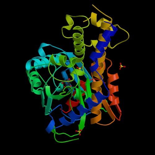
For file 2YHX:  Anderson, C.M., Stenkamp, R.E., Steitz, T.A.: Sequencing a Protein by X-Ray Crystalography. II. Refinement of Yeast Hexokinase B Co-Ordinates and Sequence at 2.1 Angstroms Resolution. J.Mol.Biol. 123 pp. 15 (1978)

For file 1BDG:  Mulichak, A. M., Wilson, J. E., Padmanabhan, K., Garavito, R. M.: The structure of mammalian hexokinase-1.. Nat Struct Biol 5 pp. 555 (1998)


Back to Main Page